
Digital Retinal Imaging & OCT Scans
Our optometrist near Hinsdale, IL use cutting-edge digital imaging technology to assess your eyes. Many eye diseases, if detected at an early stage, can be treated successfully without total loss of vision. Your retinal Images will be stored electronically. This gives your local eye doctor a permanent record of the condition and state of your retina.
This is very important in assisting your optometrist to detect and measure any changes to your retina each time you get your eyes examined, as many eye conditions, such as glaucoma, diabetic retinopathy and macular degeneration are diagnosed by detecting changes over time.
The advantages of digital imaging include:
- Quick, safe, non-invasive and painless
- Provides detailed images of your retina and sub-surface of your eyes
- Provides instant, direct imaging of the form and structure of eye tissue
- Image resolution is extremely high quality
- Uses eye-safe near-infra-red light
- No patient prep required
Marco TRS-5100―Total Refraction System
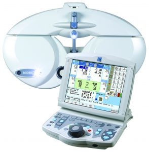 Our Oakbrook eye clinic is thrilled to announce that we now use the very latest phoropter (also called a refractor). This is the device that our eye doctor uses to assess your visual acuity and prescription needs. This latest system instantly compares your vision to your existing prescription, which makes the exam a lot easier and quicker, as well as more comfortable. With this latest optometric technology, the days of "which is better, 1 or 2?" are over. Your Oakbrook eye doctor is committed to investing in the latest equipment in order to make absolutely sure that your prescription is as accurate as possible.
Cirrus OCT―Optical Coherence Tomography
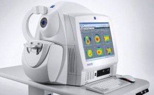 Cirrus OCT- Optical Coherence Tomography is a technique for obtaining sub-surface images of translucent or opaque materials at a resolution equivalent to a low-power microscope. It is effectively ‘optical ultrasound’, imaging reflections from within tissue to provide cross-sectional images.
This new technology is an essential instrument in the diagnosis and treatment of Macular Degeneration, Glaucoma and Diabetes.
An OCT scan is a noninvasive, painless test. It is performed in about 10 minutes right in our office. Feel free to contact our optometry practice near Hinsdale, IL to inquire about an OCT at your next appointment.
Kowa Fundus Camera
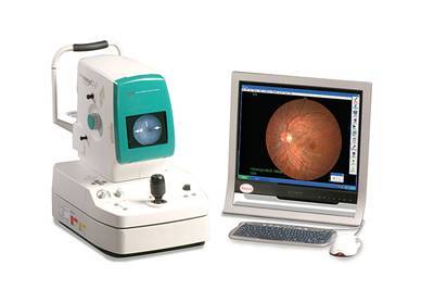
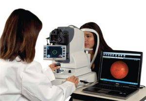 A fundus, or retinal camera, is a specialized microscope with an attached camera designed to photograph the central retina, optic disc and macula.
The resulting photograph is a high resolution magnified image which can help to assess for eye conditions including glaucoma, diabetic retinopathy, macular degeneration, optic nerve disease, and many others. This picture also gives us a baseline to compare the state of your eye health in the future.
Visual Field Testing
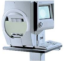
 A visual field test measures the range of your peripheral or “side” vision to assess whether you have any blind spots (scotomas), peripheral vision loss or visual field abnormalities. It is a straightforward and painless test that does not involve eye drops but does involve the patient's ability to understand and follow instructions.
The Visual Field Analyzer measures the point-by-point sensitivity of peripheral vision. This is important in diagnosing and monitoring glaucoma and neurological disorders that may have visual consequences. Each eye is tested separately and the entire test takes 15-45 minutes. These machines can create a computerized map out your visual field to identify if and where you have any deficiencies.
At our Oak Brook optometry practice, Dr. Joseph Franceschini has been serving Chicago and the Western Suburbs, including Hinsdale, for more than 20 years.
